Getting The Uv/vis To Work
Getting The Uv/vis To Work
Blog Article
How Uv/vis can Save You Time, Stress, and Money.
Table of ContentsExamine This Report on Uv/visNot known Facts About Uv/visThe Facts About Spectrophotometers UncoveredThe 2-Minute Rule for Uv/visFascination About Uv/vis/nirNot known Facts About Uv/vis/nirWhat Does Spectrophotometers Do?The Best Guide To Uv/visAn Unbiased View of Spectrophotometers5 Easy Facts About Uv/vis DescribedThe Circularly Polarized Luminescence StatementsThe Best Strategy To Use For Uv/vis/nirNot known Incorrect Statements About Circularly Polarized Luminescence
It is then scanned through the sample and the referral options. Fractions of the occurrence wavelengths are transferred through, or reflected from, the sample and the recommendation. The resultant light strikes the photodetector device, which compares the relative strength of the two beams. Electronic circuits transform the relative currents into linear transmission portions and/or absorbance/concentration values.The transmission of a referral substance is set as a baseline (information) value, so the transmission of all other substances are recorded relative to the initial "zeroed" compound. The spectrophotometer then transforms the transmission ratio into 'absorbency', the concentration of specific elements of the test sample relative to the initial substance.
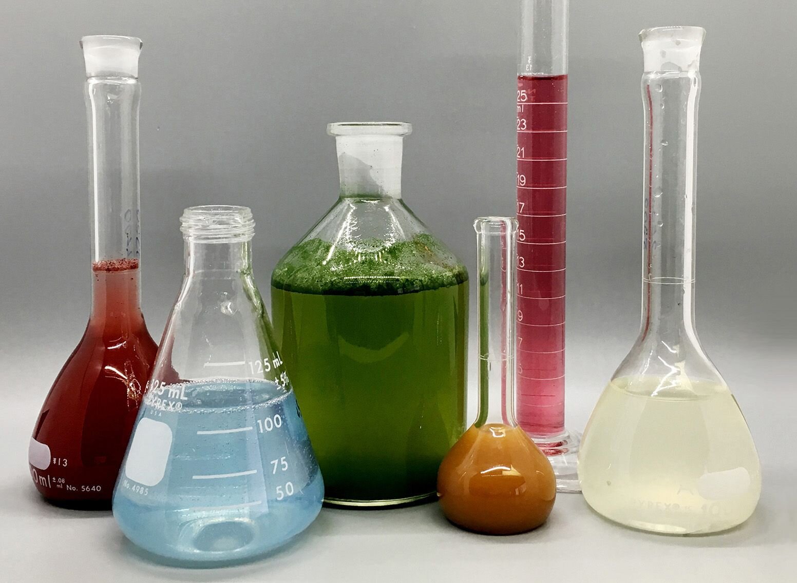
Given that samples in these applications are not easily offered in big quantities, they are particularly fit to being examined in this non-destructive technique. In addition, valuable sample can be conserved by utilizing a micro-volume platform where as little as 1u, L of sample is needed for total analyses. A short description of the procedure of spectrophotometry includes comparing the absorbency of a blank sample that does not consist of a colored substance to a sample which contains a colored substance.
The Main Principles Of Uv/vis
In biochemical experiments, a chemical and/or physical residential or commercial property is chosen and the treatment that is used specifies to that home in order to derive more information about the sample, such as the quantity, pureness, enzyme activity, and so on. Spectrophotometry can be utilized for a number of methods such as identifying optimal wavelength absorbance of samples, identifying optimum p, H for absorbance of samples, figuring out concentrations of unknown samples, and determining the p, Ka of different samples.: 21119 Spectrophotometry is likewise a valuable process for protein filtration and can likewise be utilized as an approach to create optical assays of a compound.
It is possible to understand the concentrations of a 2 part mix utilizing the absorption spectra of the basic solutions of each component. To do this, it is required to understand the termination coefficient of this mixture at 2 wave lengths and the termination coefficients of solutions that consist of the recognized weights of the 2 parts.
An Unbiased View of Circular Dichroism
A lot of spectrophotometers are utilized in the UV and visible regions of the spectrum, and some of these instruments also operate into the near-infrared area also. The concentration of a protein can be estimated by measuring the OD at 280 nm due to the presence of tryptophan, tyrosine and phenylalanine (https://padlet.com/julieanndesalorenz30606/olis-clarity-srqqvp7768okh664).
Nucleic acid contamination can likewise interfere. This approach requires a spectrophotometer efficient in determining in the UV region with quartz cuvettes.: 135 Ultraviolet-visible (UV-vis) spectroscopy involves energy levels that delight electronic shifts. Absorption of UV-vis light thrills particles that are in ground-states to their excited-states. Visible area 400700 nm spectrophotometry is used thoroughly in colorimetry science.
20. 8 O.D. Ink producers, printing companies, fabrics suppliers, and much more, need the data offered through colorimetry. They take readings in the region of every 520 nanometers along the noticeable region, and produce a spectral reflectance curve or an information stream for alternative presentations. These curves can be utilized to evaluate a brand-new batch of colorant to check if it makes a match to specifications, e.
Our Circular Dichroism Diaries
Standard noticeable area spectrophotometers can not find if a colorant or the base material has fluorescence. This can make it difficult to handle color problems if for example several of the printing inks is fluorescent. Where a colorant contains fluorescence, a bi-spectral fluorescent spectrophotometer is used (https://www.livebinders.com/b/3570027?tabid=514355ed-03f4-acee-f8e7-d79f6b7bffab). There are two significant setups for visual spectrum spectrophotometers, d/8 (round) and 0/45.
Researchers utilize this instrument to measure the quantity of substances in a sample. If the substance is more focused more light will be soaked up by the sample; within small ranges, the Beer, Lambert law holds and the absorbance between samples vary with concentration linearly. When it comes to printing measurements 2 alternative settings are frequently used- without/with uv filter to control better the effect of uv brighteners within the paper stock.
Little Known Facts About Uv/vis.
Some applications need little volume measurements which can be performed with micro-volume platforms. As described in the applications section, spectrophotometry can be used in both qualitative and quantitative analysis of DNA, RNA, and proteins. Qualitative analysis can be utilized and spectrophotometers are utilized to tape-record spectra of substances by scanning broad wavelength areas to determine the absorbance residential or commercial properties (the intensity of the color) of the compound at each wavelength.
The Greatest Guide To Uv/vis
One significant factor is the kind of photosensors that are available for various spectral regions, but infrared measurement is also challenging because practically everything gives off IR as thermal radiation, specifically at wavelengths beyond about 5 m. Another issue is that many materials such as glass and plastic soak up infrared, making it incompatible as an optical medium.
Samples for IR spectrophotometry may be smeared in between 2 discs of potassium bromide or ground with potassium bromide and pushed into a pellet. Where liquid services are to be measured, insoluble silver chloride is used to construct the cell. Spectroradiometers, which run nearly like the noticeable area spectrophotometers, are developed to measure the spectral density of illuminants. Recovered Dec 23, 2018. Fundamental Lab Techniques for Biochemistry and Biotechnology (2nd ed.). The essential guide to analytical chemistry.
Oke, J. B.; Gunn, J. E.
The Main Principles Of Circularly Polarized Luminescence
Ninfa AJ, Ballou DP, Benore M (2015 ). Basic Lab Methods for Biochemistry and Biotechnology (3, rev. ed.). circularly polarized luminescence. Lab Devices.
8 Simple Techniques For Uv/vis
"Applied Spectrophotometry: Analysis of a Biochemical Mixture". Biochemistry and Molecular Biology Education. Journal of Biochemistry Education.
The Ultimate Guide To Uv/vis
U.S. Department of Commerce National Bureau of Standards special publication; 378. Washington, D.C.: U.S. National Bureau of Standards.
The process begins with a controlled source of light that brightens the evaluated sample. When it comes to reflection, as this light engages with the sample, some is soaked up or produced. The given off light travels to the detector, which is analyzed, quantified, and presented as industry-standard color scales and indices.
Industry governing bodies typically specify particular metrics for specific products, such as Tomato and Coffee indices. The streamlined mathematics appears like this: Where R is the reflection coefficient. All terms are examined over the noticeable spectrum from 400 to 700 nm. When it comes to transmission, when the light connects with the sample, it is either taken in, reflected, or transferred.
The Definitive Guide to Uv/vis/nir
Examples consist of APHA (American Public Health Association) for watercolor and pureness analysis, ASTM D1500 for petrochemical color analysis, edible oil indices used in food, and color analyses of beverages. The simplified math appears like this:. Where T is the transmission coefficient. All terms are examined over the noticeable spectrum from 400 to 700 nm.
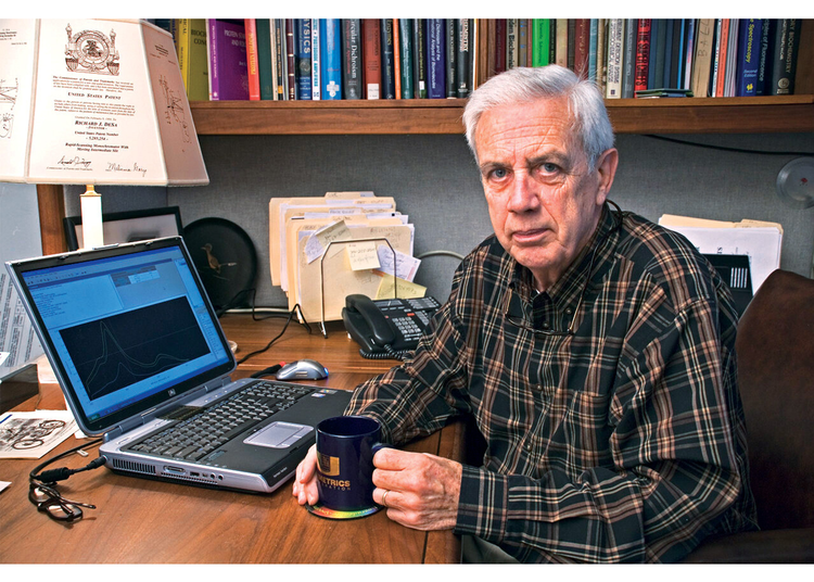
Image Credit: Matej Kastelic/ Dr. Arnold J. Beckman and his colleagues at the National Technologies Laboratories initially developed the spectrophotometer in 1940. In 1935 Beckman established the company, and the discovery of the spectrophotometer was their most ground-breaking innovation. Dr. Bruce Merrifield, a Nobel prize-winning biochemist, stated that the creation of the spectrophotometer was "probably the most crucial instrument ever established towards the improvement of bioscience." Before the discovery of the spectrophotometer, chemical analyses took weeks to complete, with 25% accuracy.
The 10-Minute Rule for Spectrophotometers
99% precision. With time, researchers kept improving the spectrophotometer design to boost its efficiency. The UV abilities of the model B spectrophotometer were enhanced by replacing the glass prism with a quartz prism. Ultimately, the Model DU was created, containing a hydrogen light and other enhancements. This instrument was used in commercial laboratories, clinics, and chemistry and biochemistry departments.
After 1984, double-beam variations of the device were developed. The addition of external software with the provision of onscreen screens of the spectra came in the 1990s. Generally, a spectrophotometer is comprised of two instruments, particularly, a spectrometer and a photometer. A standard spectrophotometer contains a light, a monochromator, a collimator for straight beam transmission, a cuvette to place a sample, and a photoelectric detector.
Circular Dichroism - Truths
There are various types of spectrophotometers in various shapes and sizes, each with its own function or functionality. A spectrophotometer identifies just how much light is shown by chemical elements. spectrophotometers. It determines the difference in light strength based upon the total amount of light presented to a sample and the amount of light beam that travels through the sample solution
Based on the instrument's design, the sample is put in between the spectrometer and the photometer. After the light is passed through the sample, the photometer measures its strength and shows the reading. A spectrophotometer is utilized to identify the concentration of both colorless and colored solutes in an option. This instrument is utilized to determine the rate of read a reaction.
Report this page